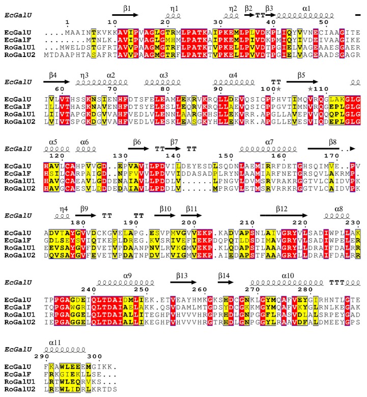
Figure 1.
Alignment of RoGalU1 and RoGalU2 with the GalU and GalF amino acid sequences of E. coli K-12 [19], executed with the MUSCLE algorithm [45,46] in the program MEGA X [47] for multiple sequence alignment with default settings and imaged with ESPript 3.0 [48]. Conserved amino acids are color-coded. Red box with white letters: strictly identical amino acids, yellow box with black bold letters: similar amino acids, which are conserved among at least two sequences, black squiggles with α or η signs: α-helical structures, black arrows with β sign: β-sheet structures, TT: strict β-turns, TTT: strict α-turns, grey stars: residues with alternate conformations.

