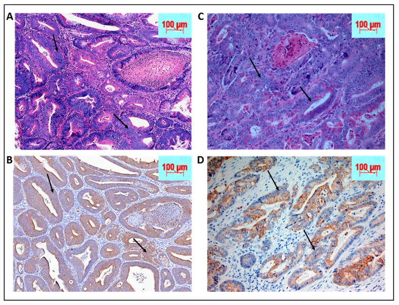
Figure 4.
(A) Biopsy of KPC: APC animals post tamoxifen induction by hematoxylin and eosin (H&E) staining at 40× magnification while (B) shows the corresponding sections stained with pan-cytokeratin to confirm the epithelial origin of the poorly differentiated tissues. On the right panel are human biopsies obtained from Montefiore medical center repository with similar staining. (C and D) Similarities in tissue morphology of the cancer between KPC: APC animal model and human. The arrows indicate some of the many tumor regions.

