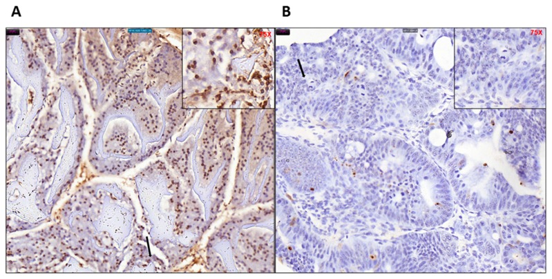
Figure 8.
TUNEL staining of (A) untreated and (B) and treated mice colon tissues showing overall less (brown) staining in the tamoxifen-treated tissue. Difference in morphology is also evident as the untreated tissue retains normal colonic architecture while the tamoxifen treated tissue is inflamed with aggregation of tumor cells, indicated by black arrow (40× magnification, 75× inset).

