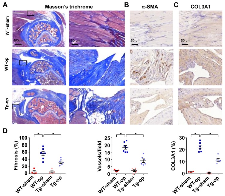
Figure 4.
Histological analysis of rotator cuff injury in wild-type mice and miR-29aTg mice. Injured tendon in miR-29aTg mice showed moderate response to the tenotomy-mediated fibrotic tissue formation (blue) as evident from Masson’s trichrome staining (A). Boxes stand for selected regions of interest for high-power field images shown in right panels. The injured sites in miR-29aTg mice showed weak α-SMA immunostaining (B) and COL3A1 immunostaining (brown) (C). miR-29a overexpression significantly improved fibrotic tissue area, vessel number and COL3A1 production (D). Data are expressed as mean ± SEM from 5–7 mice analyzed using ANOVA test and Bonferroni post-hoc test. Asterisks * indicate significant difference between groups (p < 0.05).

