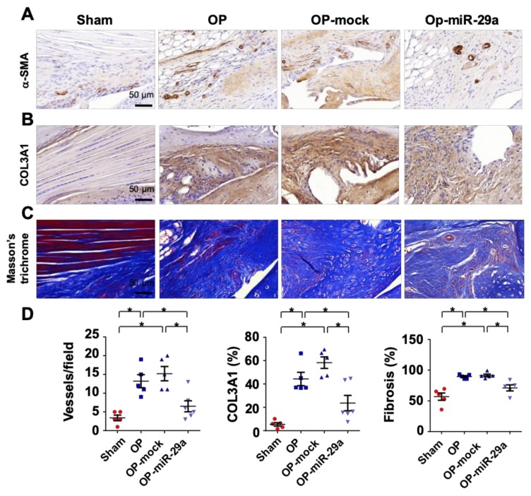
Figure 6.
Histopathological analysis of injured shoulders with or without miR-29a precursor treatment. Injured sites in the miR-29a-treated group showed moderate α-SMA immunostaining (brown) (A) and COL3A1 immunoreaction (brown) (B) along with mild fibrosis tissue formation (blue) as evident from Masson’s trichrome staining (C). The tenotomy upregulation of vessel formation, COL3A1 production and fibrotic tissue area were compromised upon miR-29a precursor treatment (D). Data are expressed as mean ± SEM from 5–6 mice analyzed using ANOVA test and Bonferroni post-hoc test. Asterisks * indicate significant difference between groups (p < 0.05).

