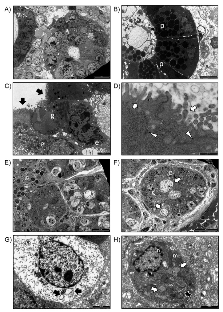
Figure 4.
(A) TEM analysis of intestinal biopsies isolated from WT mice. Villi in transversal sections (bar: 20 μm). (B) Transversal section of an intestinal villus. Paneth cells (p) were observed (bar: 20 μm). (C) Goblet cells (g) and enterocytes (e) with basally located nuclei and microvilli (black arrows) on the apical surface of the cells were observed (bar: white arrows) (bar: 500 nm). (D) Detail of microvilli on the apical surface of enterocytes (white arrows) and desmosomes on the lateral surface of enterocytes (arrowheads) (bar: 500 nm). (E) TEM analysis of intestinal biopsies isolated from Winnie mice. Transversal section of intestinal villi (bar: 20 μm). (F) Detail of an intestinal villus in which enterocytes (e) and distended cells were observed (bar: 20 μm). (G) Distended cell containing several vacuoles (black arrows) (bar: 2000 nm). (H) Enterocyte showing a preserved nucleus (n), dilated RER (white arrows), several mitochondria (m), and enlarged Golgi complex (black arrow) (bar: 2000 nm).

