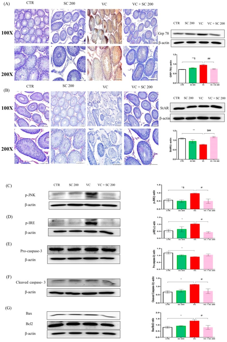
Figure 4.
SC extract upregulates protein expression levels of markers of ER stress, apoptosis, and StAR protein in VC-induced testis tissues of male SD rats. (A) Grp 78 determined by Western blot and immunohistochemistry staining. In immunohistochemical staining, weak stains were found in the control group and VC + SC 200 group. Strong dark brown staining was found in the VC group. High expression levels of Grp 78 were detected in the VC group by Western blot. (B) StAR protein level determined by Western blot and immunohistochemistry staining. In immunohistochemical staining, strong red color stains were found in Leydig cells of the control group and the VC + SC 200 group, while weak staining was noted in the VC group. Decrease in expression of StAR protein in VC was noted by Western blot. (C) Phosphorylated c-Jun-N-terminal kinase (p-JNK), (D) p-IRE, (E) pro-caspase 3, (F) Cleaved caspase 3, (G) Bax:Bcl2 ratio. Data are presented as mean ± SEM (n = 10). Statistical analyses were performed using one-way ANOVA followed by Tukey’s post hoc test. * p < 0.05 vs. CTR group, ** p < 0.01 vs. CTR group, *** p < 0.001 vs. CTR group, # p < 0.05 vs. VC group, ## p < 0.01 vs. VC group and ### p < 0.001 vs. VC group, $ p < 0.05 vs. SC group, $$ p < 0.01 vs. SC group and $$$ p < 0.001 vs. SC group. CTR, control; SC 200, SC 200 mg/kg p.o; VC, varicocele; VC + SC 200, SC 200 mg/kg; SC, Schisandra chinensis; p.o., per oral; ANOVA, analysis of variance; SEM: standard error of the mean.

