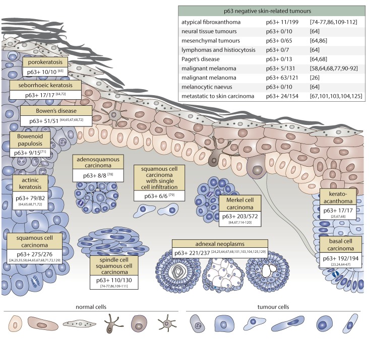
Figure 2.
p63 expression in skin cancer. p63 positively marks both benign precancerous lesions (seborrheic-, actinic-, and porokeratoses, keratoacanthoma, Bowen’s disease, and Bowenoid papulosis) and malignant tumours of the skin (basal cell carcinoma, squamous cell carcinoma, spindle cell squamous cell carcinoma (SCC), adenosquamous carcinoma, and SCC with single cell infiltration). Multiple adnexal neoplasms of both a benign and malignant character stain intensively for p63. Merkel cell carcinoma shows positive p63 expression in less than 50% of cases; indeed, its expression is correlated with poor prognosis of Merkel cell carcinoma (MCC). By contrast, neural tissue, mesenchymal tissue tumours, cutaneous lymphomas and histiocytosis, Paget’s disease, and atypical fibroxanthoma lack p63 in most cases. Metastatic carcinoma to skin stains positively for p63 in only 15% of cases. Melanocytic naevi as well as malignant melanoma are virtually all p63 negative, with the exception of one report [26].

