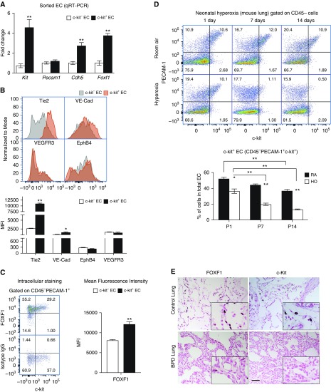
Figure 2.
Neonatal hyperoxia decreases c-KIT+ endothelial cells (ECs) in lung tissue. (A) RNA was prepared from FACS-sorted c-KIT+ and c-KIT− ECs from Postnatal Day 7 (P7) mouse lungs and analyzed by qRT-PCR. Increased mRNAs of Foxf1, Kit, and Cdh5 were found in c-KIT+ ECs (n = 3 mice in each group). Pecam1 mRNA was not changed. Expression levels were normalized to β-actin mRNA. (B) FACS analysis of P7 mouse lungs shows that c-KIT+ ECs have increased cell-surface expression of TIE2 and VE-Cadherin. Cell-surface staining of EphB4 and VEGFR3 was unaltered in c-KIT+ ECs. The mean fluorescence intensity (MFI) was calculated using n = 3 mice in each group. (C) FACS analysis shows increased amounts of intracellular FOXF1 protein in c-KIT+ ECs. Isotype control IgG was used as a control for intracellular FOXF1 staining. (D) Neonatal hyperoxia (HO) decreases the percentage of pulmonary c-KIT+ ECs in a time-dependent manner. Percentages were calculated from total number of pulmonary ECs (PECAM-1+ CD45−) using FACS analysis of wild-type lungs. Lungs of mice exposed to room air (RA) were used as controls (n = 5). *P < 0.05 and **P < 0.01. (E) Immunohistostaining shows reduced FOXF1 and c-KIT in lungs of patients with BPD (n = 7) compared with donor neonatal lungs (n = 6). Scale bar, 50 μm. BPD = bronchopulmonary dysplasia.

