Figure 4.
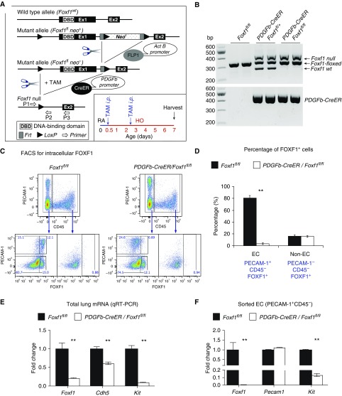
Deletion of Foxf1 from endothelial cells (ECs) inhibits c-KIT. (A) Schematic representation of the generation of PDGFb-Cre/Foxf1−/− mice and tamoxifen (TAM) treatment of newborn pups. Foxf1-floxed mice were bred with PDGFb-CreER mice to generate PDGFb-Cre/Foxf1fl/fl offspring. LoxP sites were introduced to the Foxf1 gene flanking Exon 1, containing the DNA-binding domain. TAM was given intraperitoneally at Postnatal Day 0.5 (P0.5) and P2.5. Mice were exposed to hyperoxia (HO) or room air (RA) for 7 days. (B) PCR of genomic DNA was used to identify the Cre transgene and Foxf1-null, floxed, and wild-type alleles. (C and D) FACS shows that intracellular FOXF1 staining was decreased in ECs (PECAM-1+ CD45−) but not in non-ECs (PECAM-1− CD45−), in PDGFb-Cre/Foxf1−/− P7 lungs. The percentages of FOXF1+ cells were calculated using FACS (n = 3 mice). (E and F) qRT-PCR shows that Foxf1 and Kit mRNAs were decreased in lung tissue (E) and FACS-sorted ECs (F) obtained from TAM-treated PDGFb-Cre/Foxf1−/− lungs compared with Foxf1fl/fl controls (n = 3–5 mice in each group). Expression levels were normalized to β-actin mRNA. **P < 0.01.

