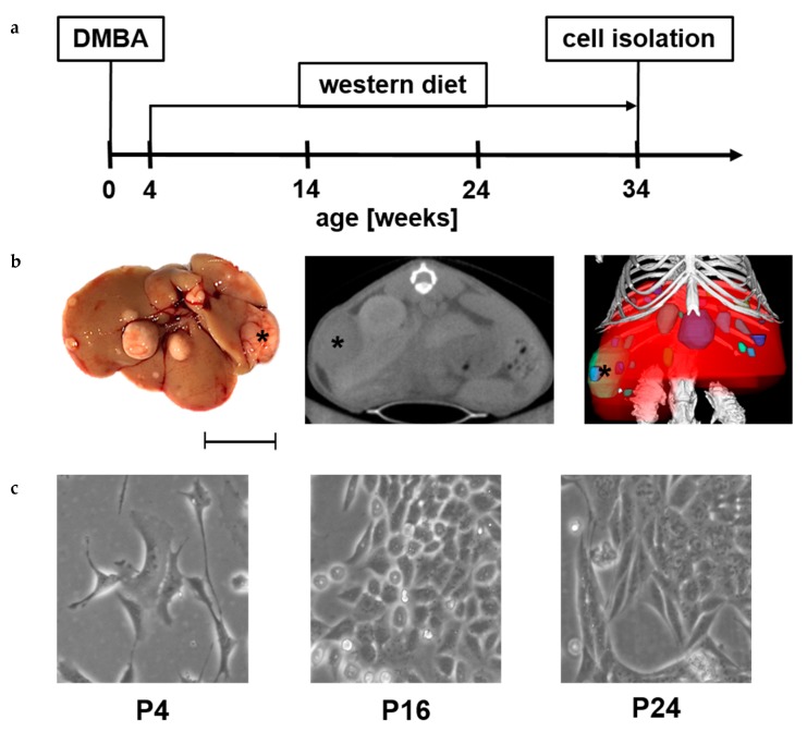
Figure 1.
Isolation of N-HCC25 cell line. (a) Male C57BL/6 mice were treated with 7,12-Dimethylbenz[a]anthracene (DMBA) on the 4th postnatal day and fed western diet for 30 weeks to develop hepatocellular cancer in NASH; (b) Characteristic picture of liver tissue that was used for isolation of N-HCC25 (view from dorsal, scale = 1 cm). Two-dimensional (2D) cross-sectional µCT image (transversal) of the mouse. 3D volume renderings of segmented bones (white), liver (red) and tumors (different colour for each tumor) upon in vivo µCT imaging (tumor marked with black asterisk, “R” marks right side of liver or mouse, scale = 1 cm); (c) Representative pictures from early and late passages of N-HCC25 (scale = 50 µm).

