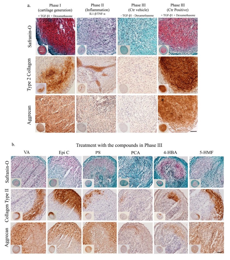
Figure 4.
Histological and immunohistochemical characterization of pellets from OA chondrocytes in the inflammatory model (long term). a) Saf-O staining, COL-II and ACAN immunostaining of pellets in three different phases as the control groups (Phase I, Phase II, Phase III). b) and after 14 days of treatment with the TCM compounds in phase III (VA, Epi C, PS, PCA, 4-HBA, 5-HMF). Scale bars = 100 μm.

