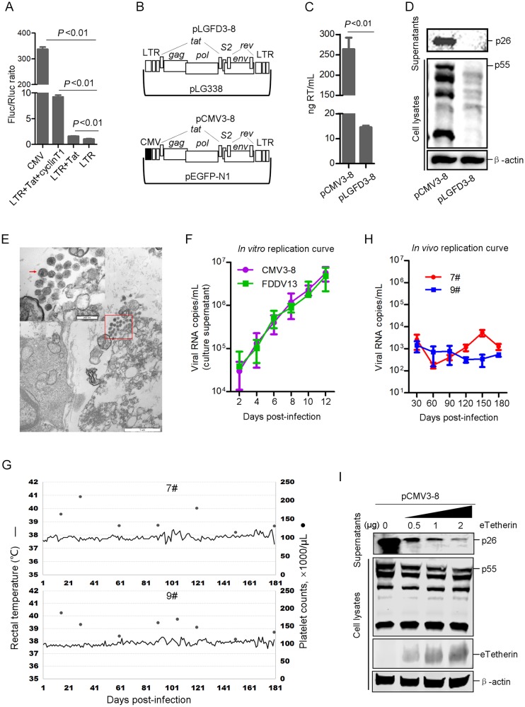
Fig. 1.
A Comparison of the transcriptional activities of the CMV promoter and the EIAV LTR without or with Tat and equine cyclin T1 in HEK293T cells. The results are the means of three independent experiments. B Schematic diagram of the EIAV infectious clones pCMV3-8 (top) and pLGFD3-8 (bottom). C Comparison of released virions from pCMV3-8- and pLGFD3-8-transfected HEK293T cells by detecting RT activity at 48 hpt. The results are the means of three independent experiments. D Comparison of EIAV expression in cell lysates and supernatants from pCMV3-8- and pLGFD3-8-transfected HEK293T cells via Western blotting at 48 hpt. The experiment was performed three times, and a representative result was shown. E TEM image of EIAV from HEK293T cells transfected with pCMV3-8 (Scale bar = 1 μm). An enlarged image of the red boxed area is shown in the top left corner (Scale bar = 200 nm), and red arrows indicate cone-shaped core virions. F Growth kinetics of the cloned virus vCMV3-8 and the parental virus FDDV13. Viral RNA was prepared from the supernatants of FDD cells infected with either vCMV3-8 or FDDV13 and was quantitatively measured by real-time RT-PCR. The results are the means of three independent experiments. (G and H) Changes in body temperature, platelet count and viral load in two horses inoculated with vCMV3-8. G The rectal temperature (━) was measured daily. An automatic blood analyzer was used to measure the platelet counts (●). H The plasma viral loads were measured by real-time RT-PCR. The results are the means of three independent experiments. I Equine tetherin-inhibited release of EIAV particles by cotransfected pCMV3-8 and eTetherin expression plasmids in HEK293T cells was validated via Western blotting at 48 hpt. The experiment was performed three times, and a representative result is shown.

