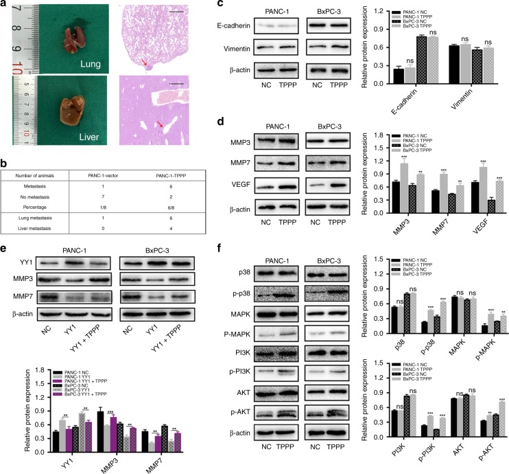
Fig. 5.
a PANC-1 vector and PANC-1-TPPP cells (1.5 × 10⁶ cells/100 μl) were separately injected into the tail vein of each mouse. Four weeks later, lung and liver metastases were evaluated by macroscopic observation and by histomorphology under microscopy. Scale bar = 200 μm. The arrows indicate the metastases. b Table listing the incidence of metastases in the nude mice treated with the vector or TPPP. c There were no statistically significant differences in the expression of EMT signalling pathway-related proteins between the two groups. d Effects of TPPP and its corresponding control group on MMP3, MMP7 and VEGF expression. e Effects of YY1, YY1 + TPPP and their corresponding control groups on MMP3 and MMP7 expression. f The expression of p38, p-p38, MAPK, p-MAPK, PI3K, p-PI3K, AKT and p-AKT in PANC-1 and BxPC-3 cells after TPPP overexpression

