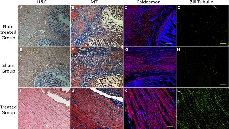
Figure 4.
Post-implantation histological analysis of IAS BioSphincters: The H&E stained surgical site section of NHP in both (A) non-treated and (B) sham group displayed irregular and discontinued arrangement of cells and extracellular matrix, but (C) 12-months post-implantation sections displayed regular arrangment of cells; MT stained sections of (D) non-treated group and (E) sham group confirmed random distribution of muscle cells (red) in collagen (blue), (F) 12-months post-implantation sections exhibited highly aligned and uniformly distributed muscle cells (red) in collagen matrix (blue); The immunostaining with caldesmon further confirmed the lack of muscle cells in both (G) non-treated group and (H) sham group, whereas (I) treated group displayed well-arranged muscle network; A poor neuronal network was appeared on immunostaining with βlll Tubulin on (J) non-treated group and (K) sham group compared to dense and innervated neuronal network in (L) treated group. Scale bar 500 µm (in A,B,E,F,I,J) and 100 µm (in C,D,G,H,K,L).

