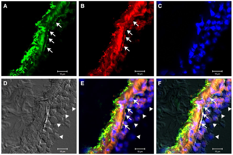
Fig. 5.

Mouse lung staining for hyaluronan (panel a, green), IαI (panel b, red), and DAPI (panel c, blue) after i.p. endotoxin exposure shows deposition of hyaluronan and IαI along the luminal endothelial surface of a venule (arrows). Panel d (transmitted light) is provided for structural reference. Panels e and f show merged images without and with transmitted light image, respectively. Note immune cells rolling along the endothelium which partially stain for IαI as well (arrowheads). Magnification: ×250
