Figure 4.
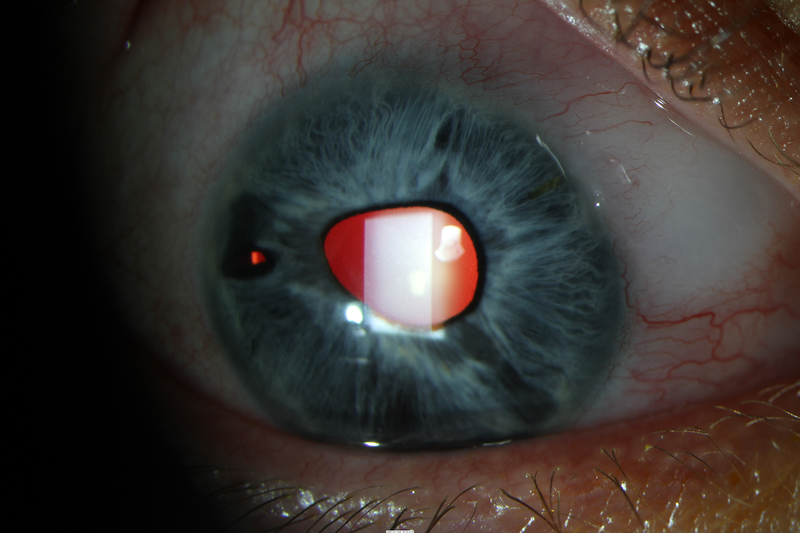
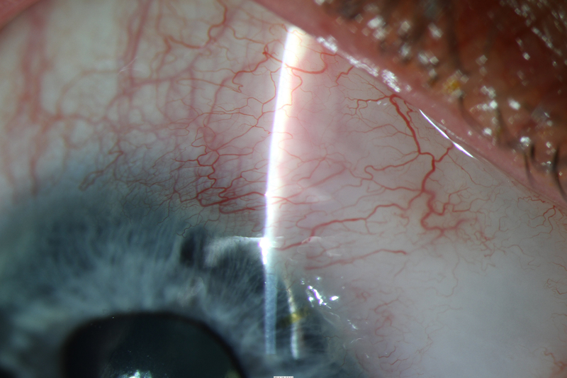
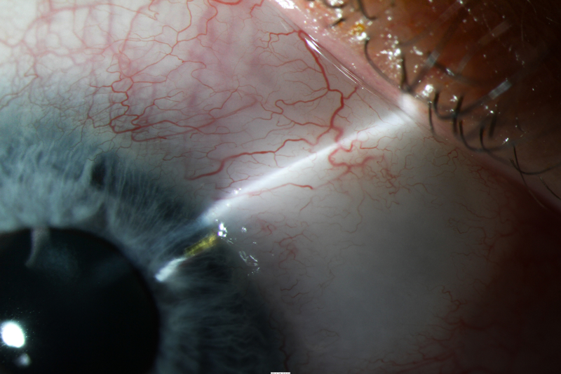
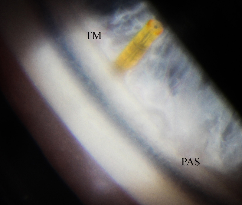
Photos of case 4 at postoperative month 5.5. A) Temporal iris atrophy and superonasal XEN bleb. B) and C) Slit lamp beams through XEN and bleb. D) Gonioscopic photograph of XEN placed such that it avoids nearby peripheral anterior synechiae. TM = trabecular meshwork. PAS = peripheral anterior synechiae.


