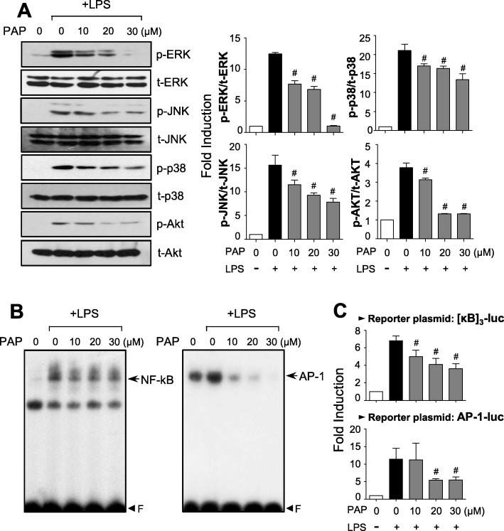
Fig. 2.
Effect of PAP on MAPKs, Akt, and NF-κB/AP-1 activities in LPS-stimulated BV2 cells. a Cell lysates were prepared from BV2 cells treated with LPS for 30 min in the absence or presence of PAP, and Western blotting was performed to determine the effect of PAP on MAPKs and Akt activity. Quantification data are shown in the right panels (n = 3). Levels of the phosphorylated forms of MAPKs and Akt were normalized to the total forms and expressed as fold changes vs. untreated control samples, which were arbitrarily set to 1. b EMSA for NF-κB and AP-1 was performed using nuclear extracts prepared from BV2 cells treated with PAP in the presence of LPS for 3 h. c Transient transfection analysis of [κB]3-luc, and AP-1-luc reporter gene activity. Data are shown as the mean ± SEM of three independent experiments. #p < 0.05 vs. LPS-treated samples

