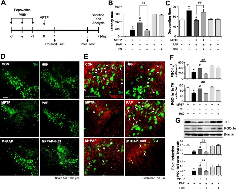
Fig. 8.
Effect of PAP/H89 on locomotor activity and the expression of TH and PGC-1α in the brains of MPTP-injected mice. a A schematic of the experimental procedure. Mice were injected with PAP (30 mg/kg, i.p.) every day for 3 days before MPTP injection. H89 was injected (1 mg/kg, i.p.) 1 h prior to every PAP injection. Mice were sacrificed 7 days following MPTP injection, and histological and biochemical analyses were performed. b, c Rotarod and pole tests were performed 1 and 6 days after MPTP injection, respectively (each group n = 12–14). d, e Immunostaining results showing TH and PGC-1α expression in the substantia nigra (each group n = 6–7). The white arrows indicate TH-positive cells with PGC-1α in their nucleus. f Quantification of TH and/or PGC-1α- positive cells in the substantia nigra pars compacta. g The protein extracts from the substantia nigra of each group were subjected to Western blot analysis using TH or PGC-1α antibodies (each group n = 5). Representative blots are provided in the upper panel, and quantification of the Western blot data is shown in bottom panel. *p < 0.05, control vs. MPTP-treated group; #p < 0.05, MPTP vs. MPTP+PAP-treated group; ##p < 0.05, MPTP+PAP vs. MPTP+PAP+H89 group

