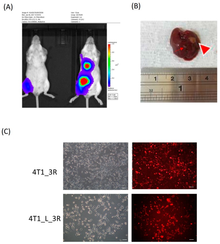
Figure 1.
Tracking and isolation of liver-metastatic 4T1_3R breast cancer cells in Balb/C mice. (A) Coronal views of bioluminescent signals acquired at different time points after initial subcutaneous (s.c.) injection of 4T1_3R cells. (B) Resection of liver for the visualization of metastatic lesions. A major lesion (big arrowhead) and a minor lesion (thin arrow) were observed in the resected liver. (C) Fluorescence microscopic examination of red fluorescent protein (RFP) expression in isolated liver-metastatic 4T1_3R cells (4T1_L_3R) compared to original 4T1_3R cells. Scale bars = 100 μm.

