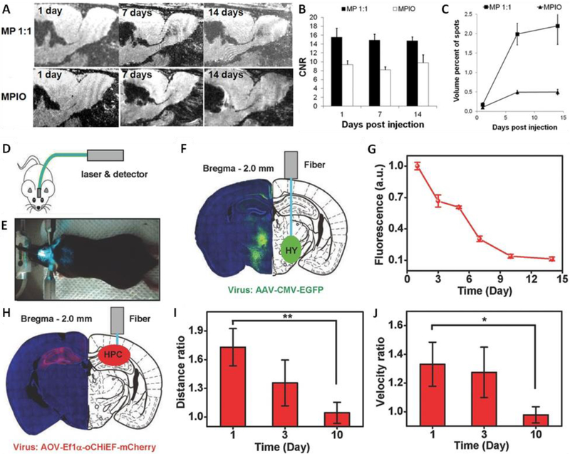
Fig. 1.
Biodegradable functional materials for neuro-imaging. (A-C) In vivo MRI of the migration of endogenous neural progenitor cells in rat brain. (A) MRI montage of same animal at level of SVZ –RMS –OB injected with microparticles (MP) or inert MPIOs. (B) CNR measurement of dark contrast within RMS. (C) Volume of dark contrast in the OB. (D-J) In vivo fluorescence photometry and optogenetic experiments with PLLA optical fibers. (D) Schematic cartoon of the experiment design and setup. E) Photographic image of the experiment setup. F) Left: confocal microscopic image of a coronal section containing EGFP. Right: schematic illustration of the fiber implanted into the HY. G) Fluorescence signals recorded via PLLA optical fibers (standard deviation, n = 6) normalized to those measured via silica fibers. H) Left: confocal microscopic image of a coronal section containing oCHiEF protein. Right: schematic illustration of the fiber implanted into the HPC. I) Ratio of travelling distance with the laser on and off. J) Ratio of travelling velocity with the laser on and off. Abbreviations: SVZ, subventricular zone; RMS, rostral migratory stream; OB, olfactory bulb; MP, microparticle; MPIO, micron sized iron oxide particle; CNR, contrast to noise ratio; EGFP, enhanced green fluorescence protein; HY, hypothalamus; HPC, hippocampus. Reproduced from Ref. [38] with permission of John Wiley and Sons.

