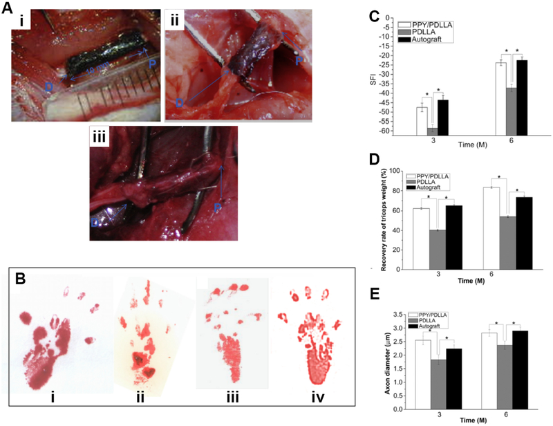
Fig. 4.
Conductive PPy/PLLA conduit for the regeneration of 10-mm sciatic nerve defects. (A) Intraoperative photographs of the PPY/PDLLA nerve conduit implantation. “P” indicates the proximal end and “D” indicates the distal end. i) Immediately after grafting. ii) 3 months postoperatively. iii) 6 months postoperatively. (B) Footprint stamps in walking track analysis after 6 months of implantation. Groups: i) PPY/PDLLA. ii) PDLLA. iii) Autograft. iv) Normal left leg. (C) Sciatic function index (SFI) as a function of implantation time. (D) Triceps weight (%) evaluation after 3 and 6 months post-operation (n= 6, * indicates p < 0.05). (E) Average axon diameter of regenerated myelinated nerve fibers. Reproduced from Ref. [41] with permission of Elsevier.

