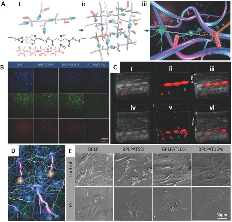
Fig. 6.
Citrate-based fluorescence/photoacoustic dual-imaging enabled biodegradable electroactive polymers. (A) i) Schematic and chemical structure of BPLPAT pre-polymers. ii) Schematic of cross-linked BPLPAT structure. iii) Schematic of functionalities of crosslinked BPLPATs. (B) Fluorescence imaging of PC12 cells uptaken with BPLP and BPLPAT nanoparticles. (C) i) Ultrasound images, ii) photoacoustic images, and iii) superimposed ultrasound and photoacoustic images of BPLP and BPLPAT scaffolds under a ~11 mm thick layer of chicken breast tissue. iv) Ultrasound images, v) photoacoustic images, and vi) superimposed ultrasound and photoacoustic images of BPLP and BPLPAT scaffolds under two layers (total ~23 mm thick) of chicken breast tissue. (D) Schematic of electrical stimulation of PC-12 cells on cross-linked BPLPAT materials. E) SEM images of PC-12 cells on BPLP and BPLPAT films without electrical stimulation (control) and with electrical stimulation. Reproduced from Ref. [36] with permission of John Wiley and Sons.

