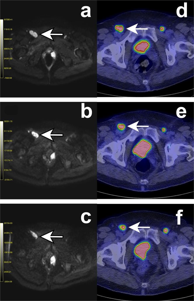
Figure 5.
DW- and PET-images of a 67-year-old woman with disseminated mucosal melanoma in vulva (cohort 2). Four intratumoral injections with AdCD40L were given to a right inguinal lymph node metastasis. The patient had partial metabolic response (PMR) in the first and stable metabolic disease (SMD) in the second post-therapy evaluation, according to EORTC criteria while the case was assessed as stable disease (SD) at both post-therapy scans according to RECIST 1.1. An increase in ADC/D in the injected metastasis was observed at both DW-MRI scans post-therapy. The short axis of the injected metastasis was measured at each time date and was unaltered according to the RECIST 1.1 criteria. An initial decrease of the f% value was observed while it was increased at the second DW-MRI scan post-therapy. In a not injected left inguinal lymph node metastasis the ADC-value was increased at both DW-MRI scans post therapy while the value of D was initially increased but it was decreased in the second post-therapy evaluation. A similar pattern as for the D-value was observed regarding the values of f% while the size of the metastasis was unaltered. A third metastasis at the proximity of the urinary bladder is present in the DW-images. This metastasis was not distinguishable at the PET/CT scans due to the high activity of the urinary bladder. (a–c) Diffusion-weighted MR image (DWI) at b = 900 s/mm2 -in the axial plane. The arrows indicate the injected right inguinal metastasis at baseline (a), in Scan 1 at week 5 (b) and in Scan 2 at week 9 (c). (d–f) FDG-PET images. The arrows indicate the injected right inguinal metastasis at baseline (a), in Scan 1 at week 5 (b) and in Scan 2 at week 9 (c).

