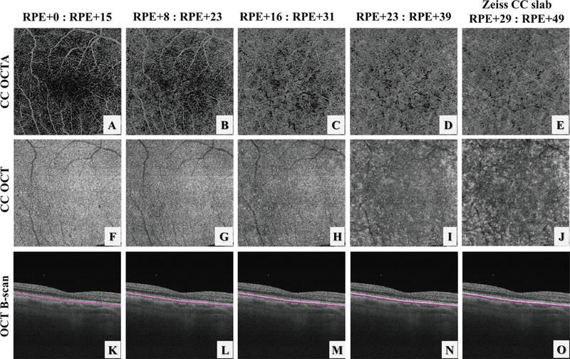
FIGURE 2.
Illustration of the choriocapillaris (CC) appearances with different selections of the CC slab position relative to the RPE in a normal eye using automatic segmentation from the instrument. (A-E) OCT angiography CC slabs with the selection of positions as shown relative to the RPE position. (F-J) corresponding OCT CC structural slabs. Note that since the axial pixel size in PLEX® Elite is 1.9 μm/pixel, the slab thickness in (D) and I is 16 μm rather than 15 μm due to rounding issues.

