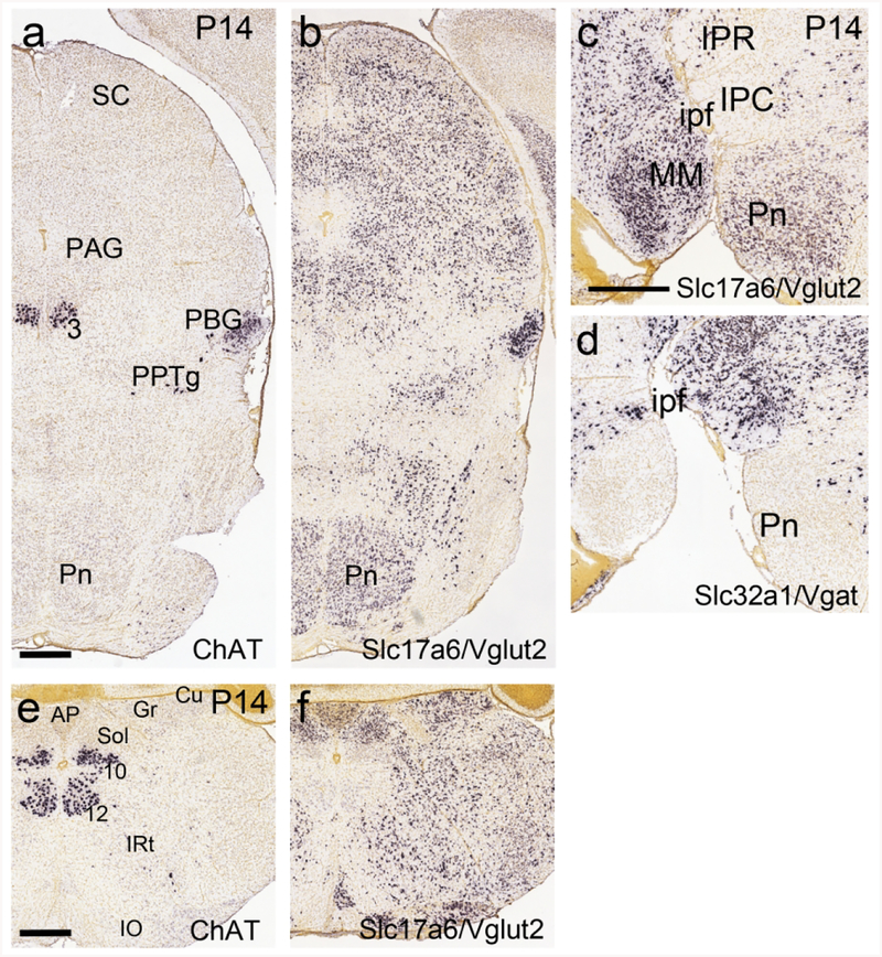
Figure 8. Extinction of ChAT expression, and expression of Slc17a6 (VGluT2), in the pontine and cuneate/gracile nuclei at postnatal day 14.
Data from the Allen Developing Mouse Brain Atlas. (a,b) Coronal section of the midbrain and pons. (a) ChAT mRNA is no longer detectable in the pontine nucleus at this stage. ChAT expression is still observed in PBG and oculomotor nucleus. (b) The pontine region is populated by glutamatergic neurons expressing VGluT2. PBG neurons appear to express VGluT2 as well as ChAT. (c,d) Sagittal sections of the pontine region, near the midline, hybridized for VGluT2 and Vgat mRNA. In (c), glutamatergic neurons expressing VGluT2 populate the pontine nucleus. In (d), few GABAergic neurons expressing Vgat are seen in the pontine nucleus, but are abundant in the adjacent IPR and IPC. (e,f) Coronal sections of the medulla hybridized for ChAT and VGluT2 mRNA. (e) ChAT mRNA is no longer detectable in the cuneate and gracile nuclei at this stage. (f) The cuneate and gracile nuclei are populated predominantly with glutamatergic neurons expressing VGluT2. Legend: 3, oculomotor nucleus; 10, motor nucleus of the vagus nerve; 12, motor nucleus of the hypoglossal nerve; AP, area postrema; Cu, cuneate nucleus; Gr, gracile nucleus; IO, inferior olive; ipf, interpeduncular fossa; IPR, interpeduncular nucleus, rostral part; IPC, interpeduncular nucleus, caudal part; IRt, intermediate reticular area (of medulla); MM, medial mammillary nucleus; PAG, periaqueductal gray; PBG, parabigeminal nucleus; Pn, pontine nucleus; PPTg, pedunculopontine tegmental nucleus; SC, superior colliculus; Sol, nucleus of the solitary tract. Scale a–b,c–d,e–f: 500μm.

