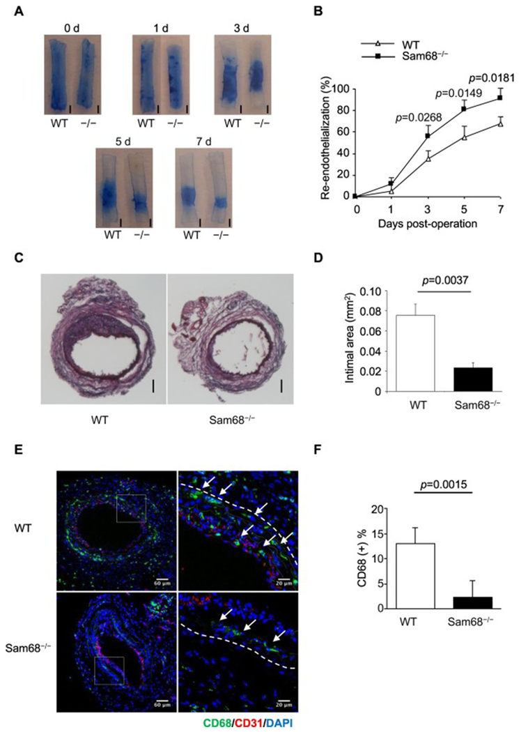
Figure 1. Deletion of Sam68 leads to accelerated re-endothelialization and attenuated neointima formation in the injured carotid artery.
Carotid denudation injury was induced by wire in the left carotid artery of WT and Sam68−/− mice. (A-B) At the indicated time points post-injury, the mice were perfused with Evan’s blue and euthanized. (A) Representative Evans blue staining, re-endothelialized areas are resistant to the blue dye and appear white (scale bar=1 mm, −/− equal to Sam68−/−). (B) Quantification of the re-endothelialized areas (n=5 per group). (C-D) At day 28 post-injury, carotid arteries were isolated and stained with Hematoxylin-Eosin. (C) Representative cross-section H.E. staining (scale bar=100 μm). (D) Quantification of neointima areas (n=5 per group). (E-F) Representative immunofluorescent CD68 staining (green staining, white arrows) (E) and quantification of infiltrating CD68+ macrophages (F) in the injured vessels at day 5 (n=5 per group). A two-tailed Student’s t-test was used in B, D, and F for statistical analysis. Error bars represent mean ± SEM.

