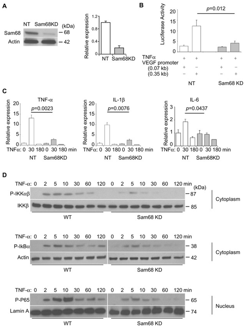
Figure 4. Knockdown of Sam68 attenuates NF-κB activation by TNF-α in macrophages.
Raw264.7 macrophages were infected with lentiviral vectors coding for Sam68-shRNA (Sam68-KD) or non-targeting shRNA (NT), and the transduced cells were selected in puromycin for 14 days. (A) Representative (left panel) and quantification (right panel) of Sam68 protein expression by Western blotting (n=4). (B) Raw264.7/NT and Raw264.7/Sam68-KD were co-transfected with NF-κB-responsive VEGF0.35 promoter-Luc or control (VEGF0.07) promoter-Luc plasmid and AP1 plasmid to allow for overnight, treated with TNF-α (10 ng/ml) for 30 min; 8 h later, the cells were lysed, and Luc activities were measured and normalized to AP1 activities (n=4 per treatment). (C) qRT-PCR analyses of cytokine expression after TNF-α treatment (10 ng/mL). n=4 per treatment per time point. (D) Western blotting analyses of phospho-IKKαβ, phospho-IKKβ, phospho-IkBα and phospho-p65 in Raw264.7/NT and Raw264.7/Sam68-KD following TNF-α treatment (10 ng/ml) for indicated times. Shown are representatives of 3-5 repetitions. A two-tailed Student’s t-test was used in B and C for statistical analysis. Error bars represent mean ± SEM.

