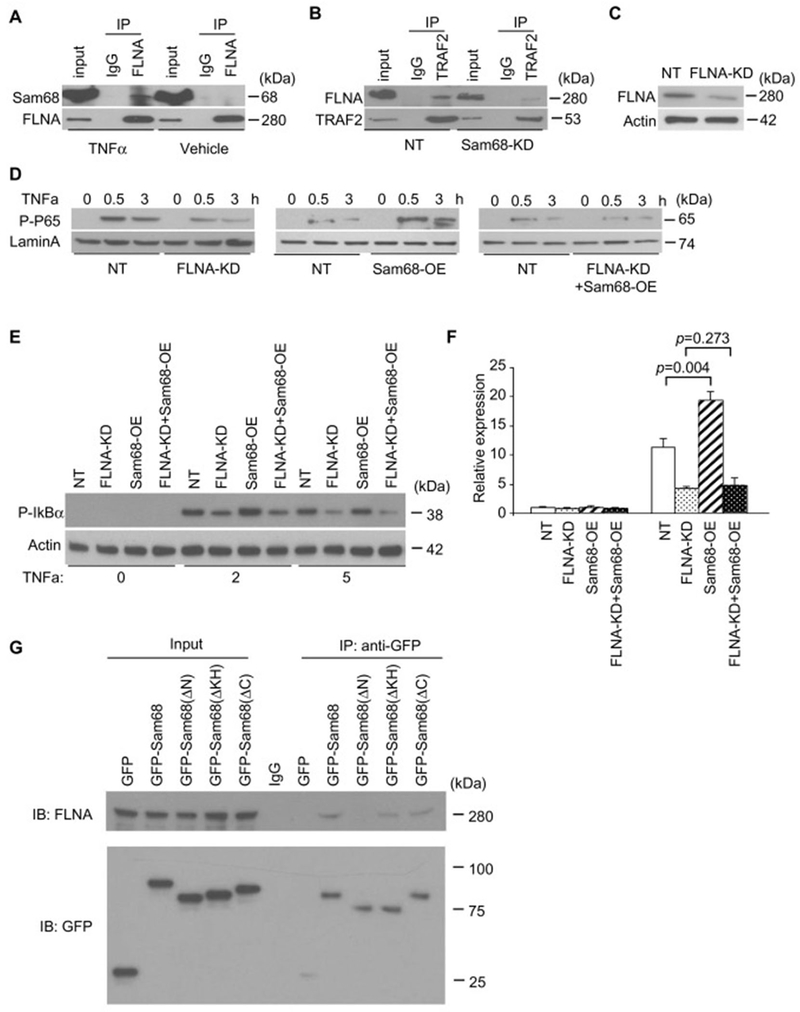
Figure 5. FLNA is a novel cofactor of Sam68 in TNF-α–induced NF-κB activation in macrophages.
(A) Raw264.7 macrophages were treated with TNF-α (10 ng/mL) or vehicle for 7 min; then co-immunoprecipitations (co-IP) were performed using anti-FLNA antibody or IgG, and the immuno-precipitates were blotted with anti-Sam68 antibody and re-blotted with anti-FLNA antibody. (B) Raw264.7/NT and Raw264.7/Sam68-KD were treated with TNF-α for 7 min; then co-IP were performed using anti-TRAF2 antibody, and the immuno-precipitates were blotted with anti-FLNA antibody and re-blotted with anti-TRAF2 antibody. (C) Raw264.7 macrophages were infected with lentiviral vector coding for FLNA-shRNA (FLNA-KD) or non-targeting shRNA (NT), then the transduced cells were selected in puromycin for 14 d, and the FLNA expression was assessed by Western blotting. (D-F) NF-κB signaling was evaluated by analyses of phospho-p65 protein (D, Western blotting), phospho-IkBα protein (E, Western blotting), and TNF-α mRNA expression (F, qRT-PCR) in Raw264.7/NT and Raw264.7/FLNA-KD cells that were transfected with Sam68 overexpression (OE) plasmid or a control plasmid and treated with TNF-α (10 ng/ml) for 0, 30, and 180 min (D) and for 0, 30 min (E-F). n=4 per treatment per time point. (G) Raw264.7 cells were transfected with GFP-Sam68 or GFP-Sam68 truncation mutants; then co-IP were performed using anti-GFP antibody, and the immuno-precipitates were blotted with anti-FLNA and re-blotted with GFP. An one-way ANOVA was used in F for statistical analysis. Error bars represent mean ± SEM.

