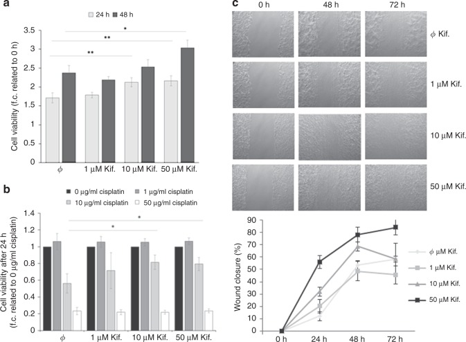
Fig. 3.
Influence of mannosidase inhibition on proliferation, platin-sensitivity and migration of ovarian cancer cells. a Proliferation of OVCAR8 cells in normal growth medium after treatment with 1, 10 and 50 µM kifunensine. Cell viability as determined by MTT test after 24 and 48 h and plotted as % values to 0 h. b Viability of OVCAR8 cells treated with 1, 10 and 50 µM cisplatin after pre-treatment with kifunensine. Cell viability was determined by MTT test after 24 h and plotted as normalised values against cisplatin untreated cells (value = 1). c Impact of kifunensine treatment on OVCAR8 cell migration ability as determined by a wound healing assay. Using ImageJ Wound Healing Tool, gap closure was quantified and represented as the percentage of cleared area remaining at 24, 48 and 72 h after the initial scratch. Representative results from one of three independent experiments (n = 3) and quantification are shown. Values are means ± s.d. *p < 0.05, **p < 0.005

