Fig. 5.
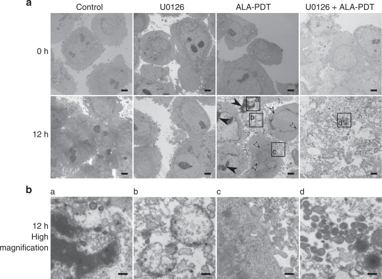
Combined MEK inhibition with 5-ALA-PDT promotes programmed cell death (PCD). a Representative low-magnification electron micrographs of DLD-1 cells at 0 and 12 h after PDT (scale bar = 2 µm). Nuclear condensation and nuclear membrane disruption are marked with black and white arrowheads, respectively. b Higher magnification micrographs of areas marked in a (scale bar = 500 nm). a nuclear condensation, b mitochondrial swelling, c cell membrane blebbing and d mitochondrial pyknosis

