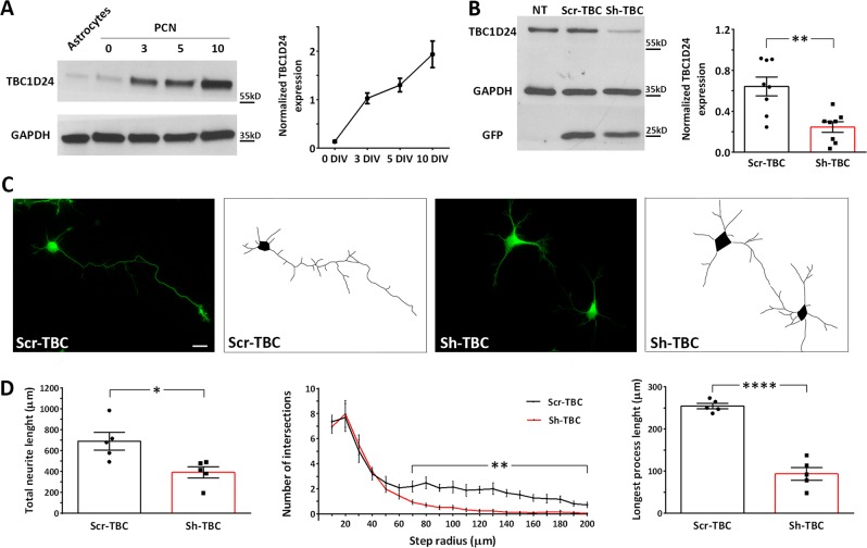
Fig. 1.
TBC1D24 expression increases in PCN at early stages of development and its silencing leads to a reduced neurite arborisation. a Representative western blot analysis showing TBC1D24 expression in rat PCNs at various stages of differentiation and in twin astrocytic cultures. Densitometric quantification with respect to time 0 is shown in the line plot on the right. Data are means ± SEM of 5 experiments from 3 independent preparations. b Representative western blot analysis showing the effective TBC1D24 silencing in rat PCNs after 5 days of transfection. Densitometric quantification is shown on the right. GAPDH is shown for equal loading and used for normalization. GFP is shown as reporter of positive transfection. Data are means ± SEM from 8 independent preparations. c Representative images of control and TBC1D24-silenced GFP-positive PCNs transfected at 0 DIV and analyzed at 5 DIV. The respective manual tracings are shown on the right of each image. Scale bar, 20 μm. d Total neurite length (left), Sholl analysis (center) and measurement of the longest neurite (right) of control and TBC1D24-silenced neurons treated as in C. Data are means ± SEM from 5 independent preparations (88 and 93 neurons were analyzed for Scr-TBC and Sh-TBC respectively). Student’t test: *p < 0.05; **p < 0.005; ****p < 0.0001

