Fig. 6.
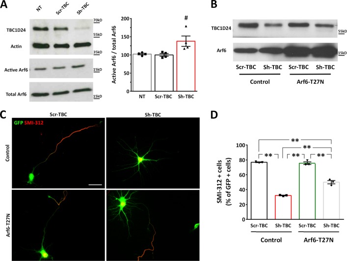
An increased Arf6 activation is responsible for the defective axonal specification in TBC1D24-silenced neurons. a Representative western blot of active Arf6 immunoprecipitation from 5 DIV rat PCNs that were either untreated (NT) or transfected with Scr-TBC/Sh-TBC. Quantification of the Arf6-GTP normalized to total Arf6 is shown on the right. Data are means ± SEM from 4 independent experiments. # p < 0.05 with One-way ANOVA/Kruskal–Wallis’ test b Representative immunoblot TBC1D24 and Arf6 expression in Scr-TBC or Sh-TBC transfected neurons (5DIV) with or without concomitant Arf6T27N overexpression. c Representative images of GFP-positive neurons transfected as above and labelled for SMI-312 at 5 DIV. Scale bar, 20 μm. d Quantification of SMI-312-positive cells with respect to the total number of GFP-positive cells from 3 independent preparations (90, 222, 202 and 224 neurons were analyzed for Scr-TBC, Sh-TBC, Scr-TBC + Arf6-T27N and Sh-TBC + Arf6-T27N, respectively). One-way ANOVA/Bonferroni’s tests: ** p < 0.001

