
Fig. 2 (abstract A63).
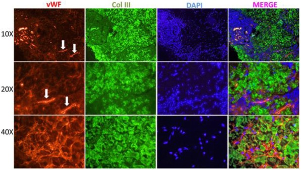
Double Immunofluorescence Staining for vWF and Collagen III in the Skin of a Patient with LS. Cuts of the reticular dermis are shown at different magnifications. There is increased collagen expression (green) throughout the reticular dermis that diffusely co-localize with increased expression of vWF (red). Vessels (arrow) stain at a higher intensity due to the presence of vWF in the subendothelium. A superficial and deep perivascular, and interstitial inflammatory cell infiltrate (blue) is also found co-localizing with vWF in areas of higher vWF expression