
Fig. 3 (abstract A63).
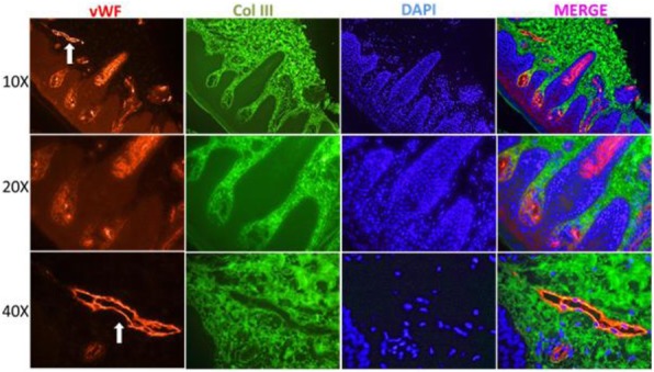
Double Immunofluorescence Staining for vWF and Collagen III in the Skin of a Patient with JDM. Cuts of the reticular and papillary dermis are shown at different magnifications. Expression of collagen III (green) is seen in the reticular dermis without co-localizing with vWF (red). Vessels (arrow) stain at a higher intensity due to the presence of vWF in the subendothelium. There is an inflammatory infiltrate at the dermal papillae and at the periphery of the vessels that co-localizes with vWF