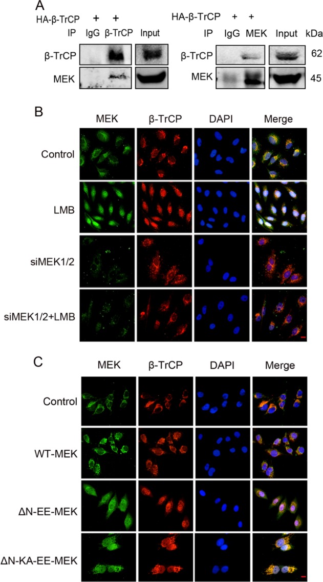
Fig. 1.

MEK nuclear translocation promotes β-TrCP localization in the nucleus. MEK localization was visualized by immunofluorescence staining with anti-MEK1/2 antibody (green). β-TrCP was shown by anti-β-TrCP staining (red). DNA was stained with DAPI (blue). Scale bar: 25 μm. a SW1116 cells were transfected with HA-β-TrCP. Anti-TrCP or anti-MEK1/2 antibody was used for IP. Blots were probed with anti-β-TrCP and anti-MEK1/2. b MDA-MB-231 cells were transfected with siMEK1/2 or negative control. Seventy-two hours after transfection, the cells were treated with LMB (10 ng/ml, 2 h) or left untreated. c MDA-MB-231 cells were transfected with control vetor, WT-MEK, ΔN-EE-MEK, ΔN-KA-EE-MEK. Seventy-two hours after transfection, the cells were treated with fresh medium containing 10%FCS for 6 h before IF
