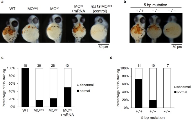
Figure 5.
(a) Significant reductions in hemoglobin (Hb) staining in the embryos were observed after rpl10a gene knockdown (MOaug and MOsp), similar to rps19 knockdown, compared to the control; embryos co-injected with rpl10a transcript recovered at 48 hpf. (b) rpl10a embryos mutated using CRISPR-Cas9 knockout showed significant reductions in hemoglobin staining. (+/+: wild-type, +/−: heterozygous, −/−: homozygous mutant). The graph displayed the percentages of normal and abnormal levels of Hb staining in (c) knocked down rpl10a gene and mRNA rescue embryos; (d) 5 bp rpl10a mutant embryos. The dense Hb staining (orange dot) at the wild-type embryo yolk sac was considered normal while a significantly pale orange or slightly colored Hb staining in the yolk sac was evaluated as abnormal. The number of animals quantified in each group are shown on top of the bars.

