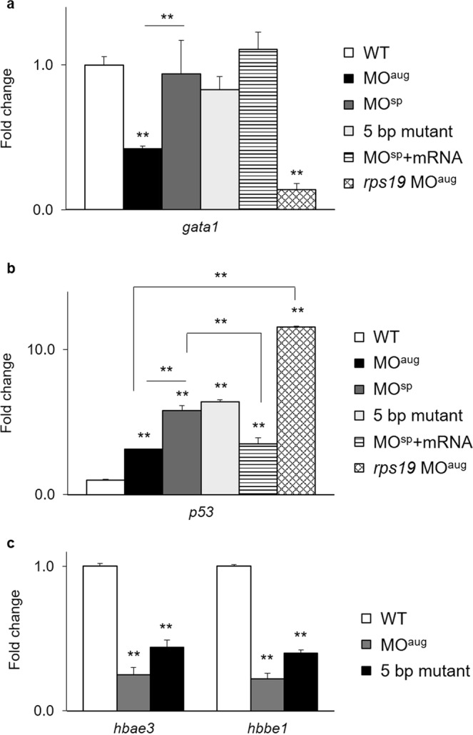
Figure 6.

Quantitative RT-PCR results showing the fold changes in the expression of the (a) gata1 and (b) tp53 genes in rpl10a MOaug, MOsp, homozygous mutant, and MOsp + mRNA injected embryos at 24 hpf compared to wild-type and rps19 knockdown embryos as controls. (c) Fold changes in the expression of hbae3 and hbbe1 mRNA in Rpl10a-deficient embryos and mutant samples at 48 hpf. Wild-type embryos were used as controls. Each group used 20 pooled embryos (replicates = 4). The data were analyzed for statistical significance by one-way ANOVA followed by Tukey’s multiple comparison test. (**p-value > 0.01).
