Fig. 2.
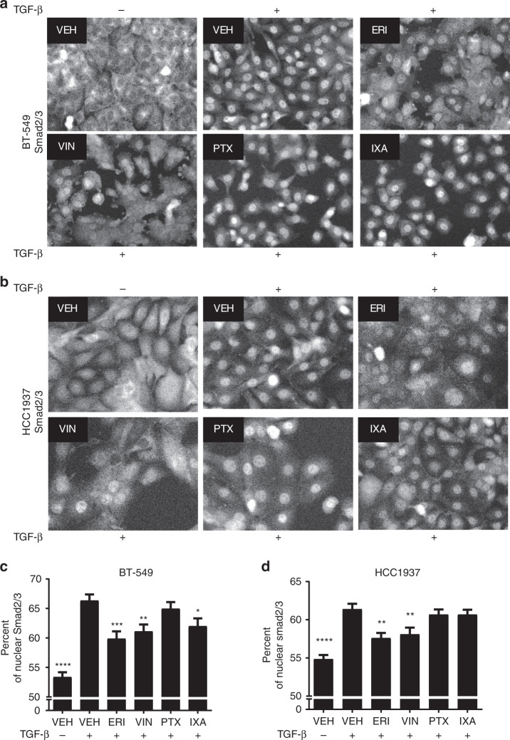
Microtubule targeting agents differentially impair the TGF-β-mediated nuclear accumulation of Smad2/3. a, b Smad2/3 localisation was evaluated by immunofluorescence in BT-549 and HCC1937 cells that were serum-starved for 18 h, pre-treated with microtubule targeting agents for 2 h, and stimulated with TGF-β for either 45 min (BT-549) or 1 h (HCC1937). Images were obtained using the Operetta™ high content imager and are representative of four independent experiments. c, d The percentage of Smad2/3 present in the nucleus of BT-549 and HCC1937 cells was quantified. Data are the average of four individual experiments, each with four replicates (mean ± SEM, N = 16). One-way ANOVA with Dunnett’s post-hoc test was used to determine statistical significance as compared to TGF-β-stimulated vehicle controls. (****p < 0.0001, ***p < 0.001, **p < 0.01, *p < 0.05)

