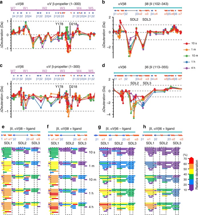
Fig. 6.
Effect of ligand binding on deuterium exchange. a–d Differences in HDX with and without saturating concentrations of TGF-β1 ligand peptide G213RRGDLATIHG223 for αVβ8 and αVβ6 are shown for each peptide plotted at the midpoint of its sequence position for a portion of the β-propeller domain and the entire βI domain. The equation for subtraction was (Dliganded − Dunliganded. Differences > 1 Da (dashed lines) are considered meaningful. All HDX data are presented in Supplementary Figs. 1 and 2. e–h Details of the ligand-binding region of the βI domain. Exchange in each peptide is colored according to the key.

