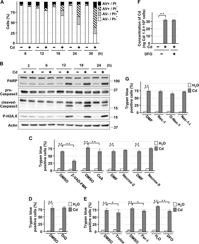
Fig. 1.
Characterization of HK-2 cell death induced by cadmium. a, b Cells were incubated with 25 μm CdCl2 (Cd) for the indicated time. Percentage of propidium iodide (PI) or Annexin-V (AV) positive cells were determined by Annexin-V and PI staining. Results are representative of at least three independent experiments a. Cell lysates were subjected to western blotting using the indicated antibodies. Immunoblots shown are representative of at least three independent experiments b. c–e, g Cells were incubated with 0.1% DMSO, 0.1% DMF, 50 μm Z-VAD-FMK, 2.5 μm CsA, 0.3 μm Necrox-2, 0.1 μm Necrox-5 c, 100 μm DiQ (d, 50 μm Trolox, 2 μm Fer-1, 100 μm DFO e, 20 μm necrostatin-1 (Nec-1), 20 μm 7-Cl-O-Nec-1 (O-Nec-1), or 20 μm necrostatin-1i (Nec-1i) g for 1 h and then incubated with or without 25 μm CdCl2 (Cd) for 30 h. The viability of cells was determined by trypan blue exclusion assay. Each value is the percentage of trypan blue-positive cells and reflects the mean ± SD of at least three experiments with duplicate assays in each experiment. f Cells were incubated with or without 100 μm DFO for 1 h and then incubated with or without 10 μm CdCl2 (Cd) for 12 h. Concentration of cadmium per 1.0 × 10⁵ cells was measured and reflects the mean ± SD of three experiments. *P < 0.05, **P < 0.01, significant difference between the samples

