
Figure 3.
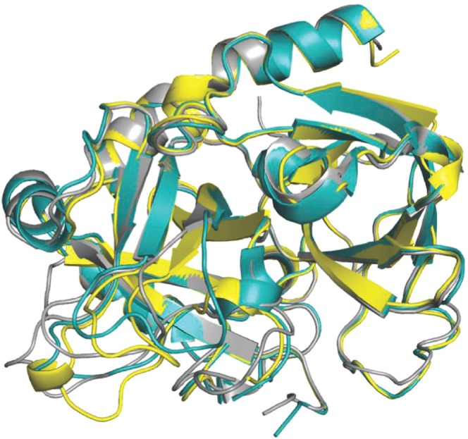
Crystal structure of the I16T mutant (yellow) aligned with the structures of thrombin in the E (rmsd = 0.39 Å; PDB entry 1SGI, grey) and E* (rmsd = 0.37 Å; PDB entry 2GP9, cyan) conformations.
Official websites use .gov
A
.gov website belongs to an official
government organization in the United States.
Secure .gov websites use HTTPS
A lock (
) or https:// means you've safely
connected to the .gov website. Share sensitive
information only on official, secure websites.
Crystal structure of the I16T mutant (yellow) aligned with the structures of thrombin in the E (rmsd = 0.39 Å; PDB entry 1SGI, grey) and E* (rmsd = 0.37 Å; PDB entry 2GP9, cyan) conformations.