Fig. 1.
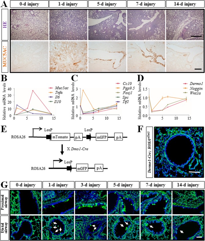
Generation of LPS injury model and Dermo1-Cre>; ROSAmTmG mice. a HE staining and MUC5AC immunostaining respectively showed the inflammatory cells infiltration and the goblet cells hyperplasia during the injury-repair process after LPS injury. b Relative mRNA levels of inflammation related genes (Tnfα, Il6, and Il10) and mucin gene (Muc5ac) were verified by qPCR. c Relative mRNA levels of different pulmonary epithelial cell marker genes (Cc10, Pgp9.5, Foxj1, Spc, and Ttf1) were verified by qPCR. d Relative mRNA levels of different pulmonary mesenchymal cell marker genes were verified by qPCR. e Schematic of Dermo1-Cre; ROSA mTmG mice. f Immunofluorescence staining showed GFP labeled pulmonary mesenchymal cells in Dermo1-Cre; ROSAmTmG mice. g Dermo1+ lineage cells emerged into the airway epithelium layer after LPS injury. In the proximal airway, GFP+ cells were clustered while in the distal airway, GFP+ cells tended to be sporadic (arrow). d, days post LPS injury. Scale bar = 50 μm

