Fig. 6.
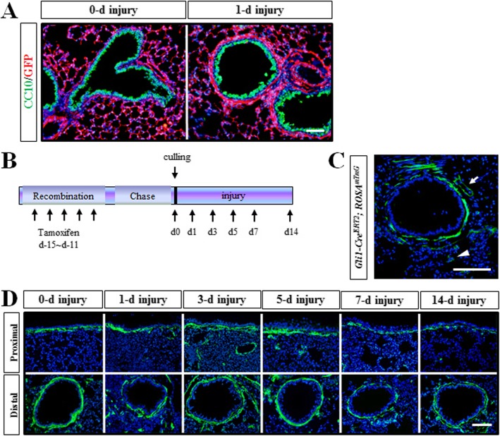
Dermo1+ stem cells were not Gli1 signaling related. a After NAPH injury, the pulmonary sub-epithelial mesenchymal layer was thickened in Dermo1-Cre; ROSAmTmG mice. b Timeline of lineage tracing of Gli1-CreERT2; ROSAmTmG-labeled cells with tamoxifen treatment in NAPH injury repair. Gli1-CreERT2; ROSAmTmG mice received five continuous dose of tamoxifen via intraperitoneal injection. Single dose of NAPH injury was conducted following 10 days of chasing. Lung tissues were collected at days 1, 3, 5, or 7. c Immunofluorescence staining results showed that GFP-labeled cells were mainly in the sub-epithelial and the peri-vascular (arrow) mesenchymal area in Gli1-CreERT2; ROSAmTmG mice. A few of GFP+ cells were also detected in the sub-meshothelial mesenchyme (arrowhead). d Gli1+ cells were detected in airway epithelium after LPS injury. d, days post NAPH injury. Scale bar 50 μm

