Fig. 1.
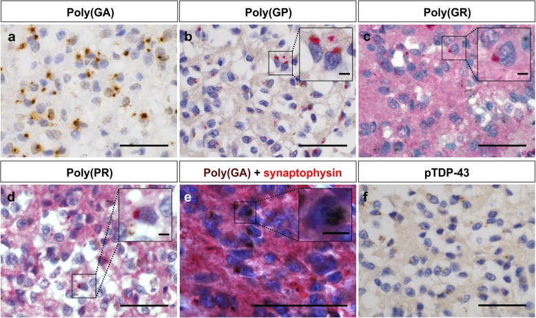
DPR and pTDP-43 pathology in the pineal gland of C9orf72 and nonC9orf72 cases. Poly(GA) (a) and poly(GP) (b) inclusions were observed in the pineal gland of all C9orf72 cases. To a lesser extent, poly(GR) (c) and poly(PR) (d) inclusions were present in the pineal gland of C9orf72 cases. Panels a-d show immunohistochemical stainings of case C9–7. The insets show a magnification of the respective DPR inclusions. The chromogen used for visualization of poly(GP), poly(GR) and poly(PR) inclusions is a Fast Red-type chromogen, therefore, a red background color was obtained. Poly(GA) inclusions (visualized by DAB) are present in the melatonin-producing pinealocytes expressing synaptophysin (visualized by Fast red) shown here in case C9–1 (e). The inset of e shows a magnification of a synaptophysin-positive neuron with a poly(GA) inclusion. The pineal gland is devoid of pTDP-43 pathology (visualized by Fast red) (f). Endogenous brown colored material was observed in the tissue (b-d, f). Scale bar represents 50 μm, the scale bar of the insets represents 5 μm. DAB stands for 3,3′-diaminobenzidine

