Fig. 4.
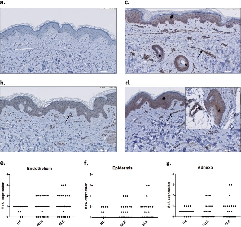
MxA expression in the skin of patients with incomplete systemic lupus erythematosus (iSLE) in comparison with healthy controls (HC) and SLE patients. Representative MxA stainings of a healthy control (a), an iSLE patient with positive MxA staining (++) of endothelium only (b), and an iSLE patient with positive MxA staining (++) of both the endothelium, the epidermis, and a hair follicle (c), and an SLE patient with positive (+++) MxA staining of the endothelium (→), the epidermis (#), a hair follicle, and sweat gland (*) (d). Dot plots of semiquantitive MxA expression in the skin of all groups are shown for the endothelium (e), epidermis (f), and adnexa (g)

