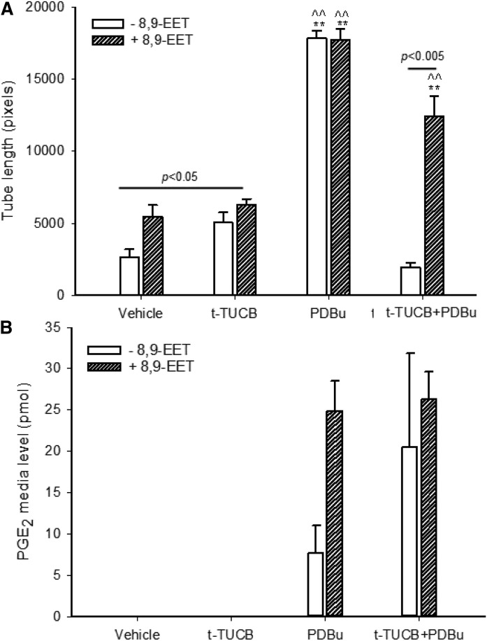
Fig. 2.
COX-2 induction, partly driven by PGE2, enhances tube formation. A: HAECs in basal media (in the absence of serum and VEGF) were seeded onto 15-well µ-angiogenesis plates with growth factor-reduced Matrigel. Cells were then treated with vehicle or 8,9-EET (0.1 µM) in the presence and absence of the sEH inhibitor t-TUCB (1 µM), COX-2 inducer PDBu (1 µM), or t-TUCB and PDBu combined. Tube length was measured manually using Fiji. Values are means ± SEs (n = 4). B: HAEC cells (300,000) were incubated with and without 8,9-EET (0.1 µM) with full media (3 ml) with and without the sEH inhibitor t-TUCB and COX-2 inducer PDBu for 20 h. PGE2 levels (pmol) in media were analyzed using LC/MS/MS. Values are means ± SEs (n = 2–3). **P < 0.001 versus the vehicle control with and without 8,9-EET; ^^P < 0.001 versus the t-TUCB treatment with and without the 8,9-EET. Statistical tests were performed using one-way ANOVA with Holm-Sidak pairwise analysis. The raw data used for this figure are reported in supplemental Table S1.

