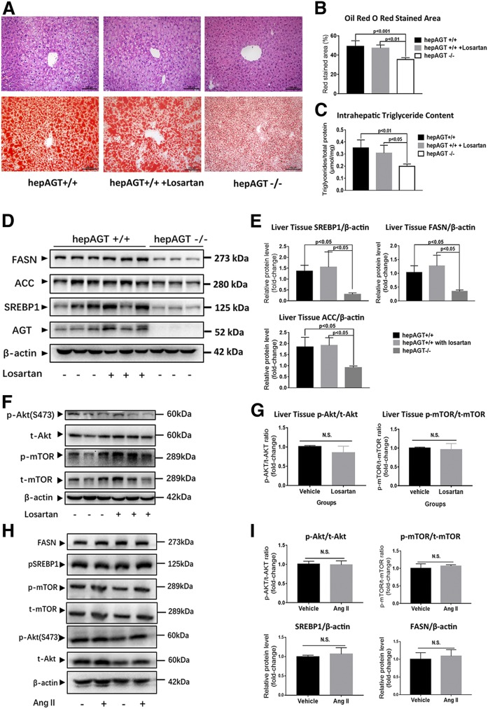
Fig. 6.
Hepatocyte-derived AGT promoted Akt/mTOR/SREBP-1c pathway activation in an Ang II-independent manner. A: The representative images of liver tissue section H&E (upper) and Oil Red O staining (lower) of hepAGT+/+ mice, hepAGT−/− mice, and hepAGT+/+ mice that received losartan administration. All of these mice were fed a Western diet for 12 weeks. B: Quantification of the percentage of red-stained area in liver tissue section Oil Red O staining (N = 5 for each group). Comparison among groups by one-way ANOVA, Holm-Sidak post hoc test. C: Quantification of intrahepatic triglyceride contents of hepAGT+/+ mice, hepAGT−/− mice, and hepAGT+/+ mice that received losartan administration (N = 5 for each group). Comparison among groups by one-way ANOVA, Holm-Sidak post hoc test. D, E: Western blotting revealed that losartan administration could not alter liver SREBP1, FASN, and ACC protein abundance in hepAGT+/+ mice fed a Western diet (N = 3 for each group). Comparison among groups by one-way ANOVA, Holm-Sidak post hoc test. F, G: Western blotting revealed that losartan administration could not alter liver Akt/mTOR signaling in hepAGT+/+ mice fed a Western diet (N = 3 for each group). Comparison between genotypes by Student’s t-test. H, I: In vitro Ang II (100 nM) stimulation exerted no significant effect on Akt/m-TOR signaling and SREBP1 and FASN protein abundance in mouse primary hepatocytes. Primary hepatocytes were incubated with 0.5 mM of palmitic acid in DMEM containing 10 nM of insulin with or without 100 nM of Ang II for 6 h (N = 3 for each group).

