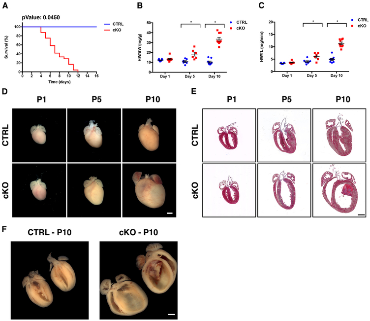
Figure 1. Loss of NEXN in cardiomyocytes results in a progressive dilated cardiomyopathy.
(A) Kaplan-Meier survival curves for NEXN CTRL (n = 35) and cKO (n = 24) mice; (B, C) Graphs showing (B) HW/BW (n=7-9 mice per time point) and (C) HW/TL (n=5-6 mice per time point) in CTRL and cKO mice. (*) Statistically significant differences with P value < 0.05. (D) Representative whole heart images from postnatal day 1 (P1), 5 (P5) or 10 (P10) mice. (E) Representative 4-chambers view Masson’s trichrome staining images of longitudinal histological sections from the same stages. (F) Butterfly cut gross anatomy showing the tridimensional organization of the organized thrombus in the left ventricle of the cKO heart and the relative CTRL. (D-F) Scale bars 1mm.

