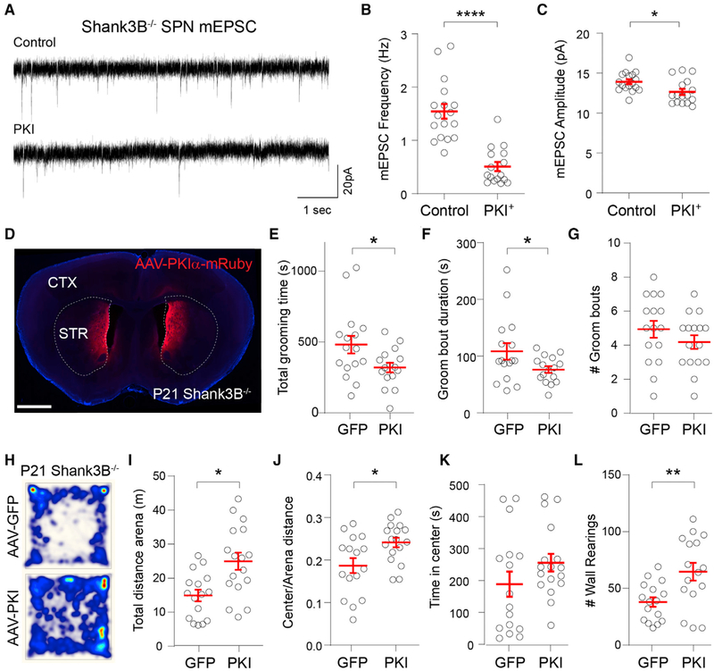
Figure 5. Postnatal Expression of PKIα in DMS Rescues Behavioral Abnormalities in Shank3B−/− Mice.
(A) Representative mESPC recordings from control or PKIα+ DMS SPNs of Shank3B−/− mice at P13.
(B and C) Mean ± SEM (B) mEPSC frequency and (C) mEPSC amplitude in control or PKIα+ DMS SPNs.
(D) Coronal section of P21 Shank3B−/− mouse infected with AAV8-Syn-PKIα-mRuby2 in DMS and ventromedial striatum (VMS) at P8. Scale bar, 1 mm.
(E) Mean ± SEM total grooming time of P21 Shank3B−/− mice expressing GFP or PKIα in the open field.
(F and G) Mean ± SEM of (F) duration and (G) number of grooming bouts per session.
(H) The total distance traveled for 20 min in the open field test by P21 Shank3B−/− mice injected with AAV-EGFP or AAV-PKIα. Heatmap represents distance normalized to AAV-PKIα.
(I) Mean ± SEM of total distance traveled in arena.
(J) Mean ± SEM of the ratio of the distance traveled in the center of the arena over the total distance traveled.
(K) Mean ± SEM of time spent in the center of the arena.
(L) Mean ± SEM number of wall rearing bouts per session.

